Anatomy of the Femoral Triangle - 3 is the Magic Number
May 23, 2022
CLICK HERE for the Interactive Femoral Triangle Quiz
Anatomy examiners tend to have favourite questions, and the femoral triangle anatomy is a firm favourite. In fact, any anatomical space comes up repeatedly in exams as they are easy questions to ask.
It is helpful to have a systematic approach when you are learning about any anatomical space. Have a series of questions that you ask yourself to jog your memory. I am going to use the femoral triangle as an example, but before I do, try clicking on the link below to try out our new 3D interactive quiz. You can spin the images around yourself
- Where is the femoral triangle located?
Start with the type of shape it is and where on the body it is located. In this example, it is a triangular region found on the anteromedial aspect of the upper thigh.
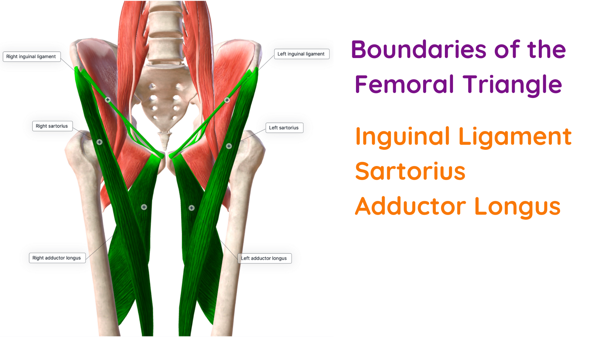
- What are the femoral triangle borders?
As it is a triangle, the femoral triangle has three boundaries. The triangle base lies superiorly, and the apex is further down the thigh. In the femoral triangle, the boundaries are formed by one ligament and two different muscles.
- superior border - inguinal ligament
- lateral border - medial edge of the sartorius muscle
- medial border - lateral edge of adductor longus
Once you know the muscles, you can be more specific. It is the medial edge of the sartorius muscle and the lateral border of the adductor longus muscle, which make up the borders of the femoral triangle.
- Which other muscles are related to the femoral triangle?
Next, define the floor or roof of the space. The floor of the femoral triangle has three muscles, and from lateral to medial, these muscles are
- iliacus
- psoas
- pectineus

- What are the femoral triangle contents?
The femoral triangle contains three main structures from lateral to medial these are
- femoral nerve
- femoral artery
- femoral vein
The femoral artery and the femoral vein lie within the femoral sheath.
I love the mnemonic NAVY, which gives you the order of the contents from medial to lateral.
N= Nerve
A = Artery
V= Vein
Y= Y-fronts!
Now you can add in more details.
Blood supply in the femoral triangle
The external iliac artery continues as the common femoral artery, which forms the superficial femoral artery and the deep femoral artery, also called the profunda femoris.
The profunda femoris gives off the medial circumflex artery and the lateral circumflex artery.
The common femoral artery gives rise to several branches within the femoral triangle.
- superficial epigastric artery
- superficial external pudendal artery
- deep external pudendal artery
- superficial circumflex iliac artery
- deep femoral artery

Venous system
Tributaries of the common femoral vein in the femoral triangle include the
- deep femoral vein
- great saphenous vein
- superficial epigastric vein
The femoral vein forms part of the deep venous system. The great saphenous vein is part of the superficial system. These two veins join at the saphenofemoral junction in the femoral triangle.

Clinical relevance
Learning anatomy just to memorise lists of words is tedious. Look for clinical details related to that space to make it more interesting. If you can get these facts out in an exam essay or a viva, you will impress your examiners big time!
You do not need much detail, but it demonstrates to the examiner that you are looking beyond the anatomy and towards the application of the anatomy.
In the femoral triangle, there are 3 clinical aspects to consider
Varicose Veins
Dilation of the great saphenous vein can result in varicose veins, a common condition characterised by large, dilated veins in the leg and calf. Varicose veins often occur due to poor valve function at the saphenofemoral junction in the femoral triangle. One of the treatment options in varicose vein surgery is tying off the great saphenous vein and then stripping the vein out.
Femoral Hernias
The femoral canal is a space found just medial to the femoral vein. It usually contains lymph vessels and nodes. In a femoral hernia, fat or a portion of the bowel pushes from the abdomen and into the femoral triangle. It is more common in women than men due to the wider-shape pelvis. Most hernias are reducible, which means they can be pushed back into the abdomen, but if the bowel becomes stuck in the femoral triangle, it can obstruct its blood supply and start to die. This is a surgical emergency, and the bowel needs to be released and put back into the abdomen as soon as possible.
Cardiac Catheterisation
Cardiologists routinely insert tubes into the femoral artery at the femoral triangle. The lines are pushed upwards to enter the heart via the aorta. Dye is then used to assess blockages in the arteries that supply the heart. Cardiac stents procedures may also be performed using this approach. Tubes are inserted into the coronary arteries to expand and increase blood flow to the heart.
Summary
If you are learning about an anatomical space, ask yourself these questions.
- Where is the space?
- What shape is the space?
- What are the boundaries?
- Does it have a roof or floor?
- What are the contents?
- Clinical features and relevance.
To make things easier, look for common themes. In the femoral triangle, it’s all about the number three.
- 3 boundaries
- 3 muscles on the floor
- 3 main contents
- 3 clinical facts
Hope that was useful – leave me a comment.
Remember to Stay Funky!
Dr Susan x
PS. Anatomy does not have to be boring. The Funky Professor has lots of content to make this easy and quick!
CLICK HERE to access all the videos and worksheets you need to pass your exams.
